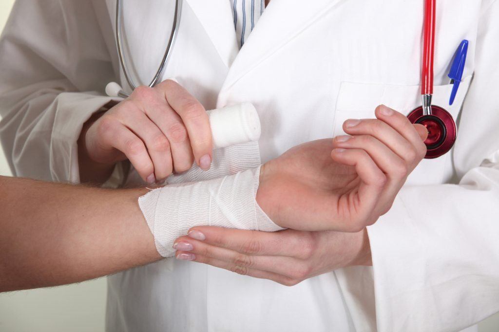
Multiple Musculoskeletal Trauma
Multiple Musculoskeletal Trauma

Multiple musculoskeletal trauma are on the rise due to the increase in extreme sports and the increase of motor vehicle accidents on the road. Musculoskeletal injuries represent more than 45 percent of inpatient rehabilitation admissions in the US.
Most high-velocity injuries result from motor vehicle accidents, motorcycle accidents, pedestrian accidents, and falls from a great height. Other causes involve occupational injuries in which the patient has been crushed between a vehicle and a solid object or has been injured by a heavy object falling on them.
Musculoskeletal injuries can involve an acute phase and a chronic phase. The acute phase involves the medical evaluation and resuscitation of the patient. Surgery is frequently required to stabilize the patient, and the patient usually needs further observation to look for other problems. The chronic phase represents the period when the patient is medically stable but requires significant rehabilitation and therapy services to gain functional capacity.
Orthopedic InjuriesThere are two types of bone, cortical bone and cancellous bone. Cortical bones are denser and have a lot of calcium in them. It is found at the ends of long bones and on bones such as the calcaneus. It also forms the outer and inner walls of the bony pelvis. Cancellous bone is softer and less structurally solid. It has a good blood supply and more bony cells. Inside the joints, there is usually smooth, articular cartilage that lines the ends of the long bones to reduce friction.
Cortical bone can withstand higher bending and compressive forces than can cancellous bone. When a cortical bone breaks, it usually has a relatively clean fracture line, although it can splinter in high-energy crashes. Cancellous bone is more likely to suffer crush or impaction type of injuries, affecting joint function.
The availability of blood supply to bones is important when determining the various options for repair and predicting whether or not the bone will heal properly. Cortical bone usually receives its blood supply from within the medullary canal, but when this type of bone fractures, the overlying blood supply around the bone increases to heal the injury. If the overlying bone (the periosteum) has been injured, the healing may be slower or may not occur at all. This can lead to more surgery to try and repair the bone. Cancellous bone has a richer blood supply from the medullary artery and does not need blood from the periosteum to heal.
Bone healing can take place by one of two methods. Primary bone healing happens when there is direct contact between the two bony segments, and there is some kind of immobilization. This can occur during plate and screw-type fixations. Secondary healing occurs when there is motion between the two fracture segments, and callus forms around the ends of the bone to help stabilize them. This is what happens with casting, bracing, and intramedullary devices.
Muscles are attached to bones using tendons. The junction between the muscles and the tendons is the weakest part, and this is the area most likely to be involved in a muscle strain. Ligaments attach bone to other bone. When the ligaments are stretched, it results in a grade one sprain. Torn ligaments that are not completely torn are called grade 2 sprains, and when a small segment of bone comes with the ligament, it is called an avulsion or grade 3 sprain.
In any injury, the bones, tendons, muscles, or ligaments can be injured. This has implications for therapy as things like range of motion and weight-bearing (or both) can be restricted, especially during the early stages of healing. Prolonged immobility can cause contractures or fibrosis, so these competing forces must be balanced for proper healing.
Methods of FixationIn today’s time, orthopedic surgeons have a wide variety of tools and techniques for treating fractures. There is rarely just one possible technique for any given type of injury. Many factors must be considered, including the patient’s age and activity level, social issues, the quality of the bone, and the presence of possible tumors or infection within the bone. The goals are to stabilize the fracture, repair or stabilize other structures, provide pain relief, and prevent future functional problems.
Casting of FracturesThe casting of fractures dates back to the ancient Egyptian period—texts from as early as the 9th and 10th centuries BC discuss plaster cast application. Modern casting now uses stronger and lighter-weight fiberglass casting. This is the type of casting used to treat children because they have excellent healing and remodeling potential.
Casts in adults are less commonly used. They only provide relative stability and don’t do well in unstable fracture patterns. They block the skin and soft tissue and are relatively heavy. Casts are used as a temporary measure in multiply injured patients before definitive surgical therapy can be undertaken. Casts are usually applied for a minimum of 4 weeks and usually longer.
Functional BracingFunctional bracing is a good alternative to casting because it provides some stability to the injured area but allows for the motion of the adjacent joints. The braces can be removed to evaluate the skin underneath and for hygiene purposes. They tend to be lighter than casts. They are often used when surgery is not warranted or in the subacute phase after surgical repair to protect the area. There are braces for the humerus, the ankle, and the knee. Weight-bearing is usually allowed with functional braces, and range of motion is usually encouraged within limitations. Custom functional braces are also used for specific regions and areas.
External FixationThis involves the use of pins or wires inserted through the skin and into the bones. The pins or wires are attached to Bares or rings to stabilize them. Temporary external fixation is usually applied when the injury pattern or the patient’s medical condition does not allow for a more definitive fixation within a few days from the injury. Soft tissue swelling, damage to the skin or underlying muscle, and the location of the injury determine whether or not external fixation is used. External fixators can be used to lengthen short bones and the correction of a bony deformity.
Whether the external fixator is on for a long time or a short time, it is always important to evaluate the sites where the pins enter the skin as these are frequent sites of infection. The infections are usually localized and are treated with oral antibiotics; however, they can progress to become deeper abscesses or osteomyelitis (infection of the bone). To prevent this, pins are put into a region where internal hardware will go later. Patients or family members can be taught the basic care of pin sites, which usually reduces the risk of pin infections. Sometimes the pins will need to be removed or switched to a different site. Weight-bearing is usually prohibited for temporary external fixators but is encouraged when long-term external fixators are used.
Intramedullary FixationIntramedullary devices are placed down the shaft of a long bone where the bone marrow usually is. They will be placed using small incisions near the hip or knee for lower extremity fractures or around the shoulder, elbow, or wrist for humerus and forearm fractures. Intramedullary fixation is most commonly used for femur fractures. These can allow for limited weight-bearing soon after surgery. Because they use small incisions, there is a fast recovery and a chance for early rehabilitation.
The most common complication of intramedullary fixation is pain around the entry site, usually around the hip or the knee. This usually occurs about 50-60 percent of the time. The pain usually goes away after about a year after surgery, although some patients will require nail removal for pain relief. The reason for this sort of pain is unclear.
Intramedullary nails usually have cross-locking screws at the top and bottom ends to prevent rotation of the fracture. They may be prominent or can cause local irritation from the movement of the tendons and muscles over their heads or tips. These screws have to be removed often after the fracture has healed because they locally irritate the tissues.
Plate Screw FixationPlates and screws are one of the most common ways to treat fractures, especially those around joints. There are specialized plates designed for a particular region and fracture type. There has also been an interest in using smaller incisions to place these kinds of plates to not to damage the blood supply.
Plates that provide rigid compression are usually used in the forearm and humerus and may allow for early range of motion and weight-bearing. Buttress-style plates are used around the joint areas, especially in ankle fractures. They do not allow for immediate weight-bearing but are usually stable enough to permit a range of motion exercises.
Joint Replacement for TraumaOn occasion, a fracture in a knee, hip, shoulder, or elbow is so severe that it would be impossible to reconstruct the fracture so that a joint replacement is necessary. These joint replacement surgeries have an excellent track record when done for arthritis but can be problematic for fractures. With fractures, there can be soft tissue injuries or a loss of bony landmarks that make the replacement surgery harder. Outcomes are often less successful. The final range of motion may be less than otherwise anticipated, and the rehabilitation usually must proceed at a slower pace. Range of motion exercises is encouraged as soon as possible following surgery. In the case of hip or knee replacement, there may be limited weight-bearing so that the replacement has a chance to heal. With elbow or shoulder replacements, the range of motion is started immediately, but weight-bearing is delayed for approximately six weeks.
Traction TreatmentTraction used to be the mainstay of treatment of lower extremity fractures, but this has largely been replaced by definitive surgery, with traction used temporarily before surgery. Femoral and pelvic fractures are the types that usually require temporary traction. However, if the patient has other medical problems, traction can be used for up to six weeks, after which casting or bracing is used for another six weeks.
The major downsides of traction are that the patient is usually bed-bound for the six-week period, and there is an increased risk for deep vein thrombosis and pressure sores. There must be a close follow-up in the first few weeks to ensure that the fracture ends are properly aligned. The traction pin sites must be monitored closely and kept clean. Physical therapy can be done on the other extremities to try and prevent atrophy of all the extremities.
Geriatric PatientsAs the number of active geriatric patients increases, there are more likely to fracture in this population. It is hard to know what constitutes geriatrics. The age of 75 years is probably a good cut-off even though certain individuals older than 75 are physiologically younger and can be treated as such. For the most part, geriatric patients tend to have more medical comorbidities and have a greater risk of surgical procedures complications. The risk of mortality is greater, even several months out from the injury. Studies have shown that an elderly person with a hip fracture has only a 50 percent chance of regaining pre-injury activities and has a threefold increase in mortality if they have other comorbidities. It is less a problem of the actual injury and more a result of the associated complications.
With fewer physiological reserves, even minor complications can have a major impact. They often can’t use a walker or crutches because of poor upper body strength or balance problems. This can lead to bedridden or wheelchair-bound, increasing the risk of pneumonia, deep vein thrombosis, or pulmonary embolism. They may be more sensitive to postoperative medications for pain, which can cause delirium or problems with balance and walk. They are more likely to have osteoporosis that can affect the stability of the repair and can put them at risk for additional fractures from even minor trauma.
Poly Trauma PatientsPolytrauma patients have at least two long bone fractures or a long bone fracture and a pelvic fracture along with injuries to other body areas, such as the abdomen or chest. These patients require a longer stay in the ICU and may require several surgical procedures over a short period of time to fully address all their injuries.
While in the ICU, they may be intubated or unable to follow commands, and beginning rehabilitation can be difficult. At the same time, they are at an increased risk of atrophy of their extremities as their body tries to heal from all of their various injuries. This means that proper nutritional support and proper positioning aids, and range of motion exercises are usually necessary.
Once the polytrauma patient has recovered from their injury and subsequent surgeries, they may be very limited in their mobility. They may have a significant weight-bearing restriction on multiple extremities. Many of these patients need admission to a skilled nursing facility to allow for some healing before participating in a more aggressive rehabilitation program. The initial rehab goals may need to be scaled back because of atrophy and impaired nutritional status.
Polytrauma patients that have suffered a traumatic brain injury can be particularly challenging. In the acute phase after their injury, they may be confused and unable to participate in rehabilitation. Depending on the severity of their injury, they may recover somewhat but still have difficulty with more complex tasks and short-term memory. The situation can be complicated when it comes to weight-bearing restrictions, or precautions for a range of motion have been imposed because the patients often attempt to get out of bed, unaware of the extent of their injuries. Traumatic brain injury is also associated with heterotopic ossification, in which injured muscle and soft tissue turn to bone. This can greatly impact a range of motion exercises, especially when it involves areas around the joint. Close follow-up and observation are necessary to detect this complication.
Watch YouTube Video: Management of Patients with Musculoskeletal Trauma. The following video provides an overview of managing patients who suffer from musculoskeletal trauma.
Editor’s Note: This page has been updated for accuracy and relevancy [cha 4.13.21]
Photo by Pixabay